Day Two ... Recording, Shaping and Dissecting!
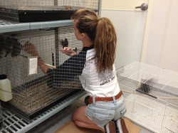
I think day two requires a preface of what Krista and I worked on on Friday. Although most of Friday was used to introduce me to everything around the lab, Krista and I were able to go grab a bird (number B767, to be exact!) for me to "shape." (Don't let yourself be confused by the terminology, most of it is pretty intuitive.) "Shaping" basically just means training the bird in a particular behavior that we've designed for it. So after the bird was set up in it's cage, I piled some food on top of the "hopper" for it, so that it would know where it's food will be coming from in the future. The "hoppers" are lift-able food dispensers for the bird - controlled by the pecking of the bird in ports above the hopper bowl itself (unless, of course, the bird isn't shaped yet, which requires you to tape the hopper up so the bird can get food, like Krista and I did for my bird 767 at first).
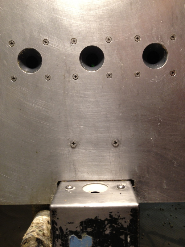
Fast-forward to Monday, May 27th, which was my second day at the lab. Bird 767 had eaten all the food off the top of the hopper, which meant he knew where his food was coming from, so Krista advised me to start manually shaping him. Manual shaping just means that I stand next to the control board, and watching three phases of the bird's behavior: 1) Standing by or on the hopper, 2) Looking at the pecking ports, 3) Pecking at the ports. When I manually shape him, I have to watch when he does these three activities in order, and lift the hopper by flicking a switch on the control board. This process is designed to first, get him used to the sound, shake and idea of the hopper and second, to get him to understand that he is rewarded with food from the hopper for pecking inside the ports. After Krista and I could see that he understood the process, we started him on a programmed shaping system run by the computer. (If you're curious what a hopper looks like, you can check out the photo on the side bar which shows the hopper bowl and three pecking ports, or you can go the "Photo Essay" page for multiple perspectives.)
Shaping wasn't the only thing Krista and I covered on our second day - we also started recording! The term "recording" is a bit counter-intuitive, because you might assume, as I did, that it refers to recording tones and motifs of birdsong. In fact, recording actually refers to recording activity in a bird's brain. All of this required unique preparations in terms of the system and for the bird as well.
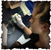
Preparation Krista and I went through in order to perform the recording began with surgery on our bird, which of course was super exciting for me! We started by giving the bird an anesthetic called isoflourane, which is generally administered through a box which is attached to an oxygen tube. In that case, the bird would be put in the box while awake, and removed when it was asleep. Krista, however, being the genius that she is, developed a faster and easier way to administer the isoflourane; she uses the same mask to anesthetize the bird as she does to keep him asleep during the procedure. During that process, she wraps the tip of a latex glove around the end of a small oxygen tube and then positions it around the bird's beak and over it's nostrils.
Once the bird was fully asleep, she attached it to a head plate that clamps around it's beak and through it's ears to hold the bird steady. Then, the surgical process began! The first step was to make sure we could get a clear look at the head of the bird - so we had to de-feather him... Which was actually more difficult that I thought it would be. Once it was de-feathered, Krista used surgical scissors to cut open the skin above the part of the skull that covers the auditory cortex of the brain (the part we care about, and would record from the following day). Once that was done, Krista used dental cement (the goop they use to attached braces to your teeth) to surround the portion of the skull that would be exposed once we shaved off the first layer of skull, as well as to attach the pin which would be used to attach and steady the bird to the recording equipment. Once the cement was laid and the pin was placed, Krista used a Dremel drill to help us reach the trebeculum layer of the skull. The trebeculum is a meshy-looking second layer of skull. For the final step of surgery, we covered the exposed part of the skull with body cast, so that the skin and skull wouldn't heal back or bleed. After we cleaned the surgical area, we brought the bird back to it's home as it started to wake up from the anesthetic. We gave him his food and water bottle back, plus some extra food and crickets in reward of the success of surgery (just like humans, the anesthetic makes the bird a bit nauseous, so his food and water is removed from his cage only for the hour before surgery).
I think day two requires a preface of what Krista and I worked on on Friday. Although most of Friday was used to introduce me to everything around the lab, Krista and I were able to go grab a bird (number B767, to be exact!) for me to "shape." (Don't let yourself be confused by the terminology, most of it is pretty intuitive.) "Shaping" basically just means training the bird in a particular behavior that we've designed for it. So after the bird was set up in it's cage, I piled some food on top of the "hopper" for it, so that it would know where it's food will be coming from in the future. The "hoppers" are lift-able food dispensers for the bird - controlled by the pecking of the bird in ports above the hopper bowl itself (unless, of course, the bird isn't shaped yet, which requires you to tape the hopper up so the bird can get food, like Krista and I did for my bird 767 at first).
Fast-forward to Monday, May 27th, which was my second day at the lab. Bird 767 had eaten all the food off the top of the hopper, which meant he knew where his food was coming from, so Krista advised me to start manually shaping him. Manual shaping just means that I stand next to the control board, and watching three phases of the bird's behavior: 1) Standing by or on the hopper, 2) Looking at the pecking ports, 3) Pecking at the ports. When I manually shape him, I have to watch when he does these three activities in order, and lift the hopper by flicking a switch on the control board. This process is designed to first, get him used to the sound, shake and idea of the hopper and second, to get him to understand that he is rewarded with food from the hopper for pecking inside the ports. After Krista and I could see that he understood the process, we started him on a programmed shaping system run by the computer. (If you're curious what a hopper looks like, you can check out the photo on the side bar which shows the hopper bowl and three pecking ports, or you can go the "Photo Essay" page for multiple perspectives.)
Shaping wasn't the only thing Krista and I covered on our second day - we also started recording! The term "recording" is a bit counter-intuitive, because you might assume, as I did, that it refers to recording tones and motifs of birdsong. In fact, recording actually refers to recording activity in a bird's brain. All of this required unique preparations in terms of the system and for the bird as well.
Preparation Krista and I went through in order to perform the recording began with surgery on our bird, which of course was super exciting for me! We started by giving the bird an anesthetic called isoflourane, which is generally administered through a box which is attached to an oxygen tube. In that case, the bird would be put in the box while awake, and removed when it was asleep. Krista, however, being the genius that she is, developed a faster and easier way to administer the isoflourane; she uses the same mask to anesthetize the bird as she does to keep him asleep during the procedure. During that process, she wraps the tip of a latex glove around the end of a small oxygen tube and then positions it around the bird's beak and over it's nostrils.
Once the bird was fully asleep, she attached it to a head plate that clamps around it's beak and through it's ears to hold the bird steady. Then, the surgical process began! The first step was to make sure we could get a clear look at the head of the bird - so we had to de-feather him... Which was actually more difficult that I thought it would be. Once it was de-feathered, Krista used surgical scissors to cut open the skin above the part of the skull that covers the auditory cortex of the brain (the part we care about, and would record from the following day). Once that was done, Krista used dental cement (the goop they use to attached braces to your teeth) to surround the portion of the skull that would be exposed once we shaved off the first layer of skull, as well as to attach the pin which would be used to attach and steady the bird to the recording equipment. Once the cement was laid and the pin was placed, Krista used a Dremel drill to help us reach the trebeculum layer of the skull. The trebeculum is a meshy-looking second layer of skull. For the final step of surgery, we covered the exposed part of the skull with body cast, so that the skin and skull wouldn't heal back or bleed. After we cleaned the surgical area, we brought the bird back to it's home as it started to wake up from the anesthetic. We gave him his food and water bottle back, plus some extra food and crickets in reward of the success of surgery (just like humans, the anesthetic makes the bird a bit nauseous, so his food and water is removed from his cage only for the hour before surgery).
 RSS Feed